Prostate Artery Embolization (PAE)

PAE is a medical procedure that began in 2012 and has since gained popularity. It is a minimally invasive medical procedure used to treat enlarged non-cancerous prostate glands. PAE uses advanced technology to treat benign prostate hyperplasia, also called BPH, a prevalent condition in aging men.

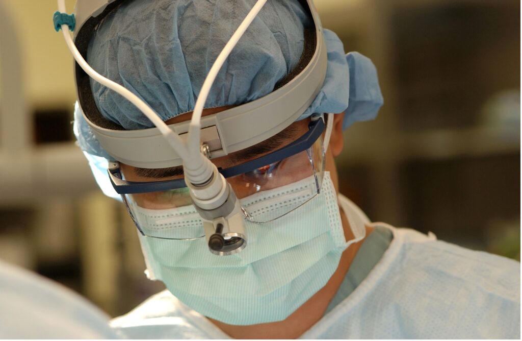
Prostate Artery Embolization reduces urinary tract symptoms caused by BPH, such as bladder and kidney problems. An Interventional Radiologist (IR) uses advanced imaging techniques to view internal body parts and issue treatment without surgery.

The Prevalence of Prostate Artery Embolization

The symptoms of PAE linked to benign prostatic hyperplasia are evident in one in four men 55 years. In the United States, almost half of men 50 years of age have a larger prostate, and 70% of men between 60-69 years and 80% aged 70+ also suffer from benign prostatic hyperplasia.

In case you are among men with such conditions, MG Health has the answers to your problems. MG has some of the best experts in PAE.

The Achievement Rate of Prostate Artery Embolization

[https://pixabay.com/illustrations/kidney-anatomy-biology-body-cancer-3667909/]

PAE boasts high success rates, with more than 90% of males getting relief in a year. On the good side, PAE has no adverse sexual performance side effects, unlike other treatments.

Some studies also indicate that PAE has lifelong success in medicating growing prostate.

A review shows that more than 80% of men are doing well three years into a PAE procedure.

Signs and symptoms of Benign Prostatic Hyperplasia

  • Difficulties in starting urinating or unable to empty your bladder
  • Presence of blood in urine, your urine may be pinkish
  • Weak urine streams, i.e., stop and start urinating suddenly
  • Dribbling at the end of urination
  • Urinary tract infections (UTIs)
  • Frequent urination, especially at night
  • Feeling the urgency to urinate all the time
  • Hematuria, bleeding from the enlarged prostate

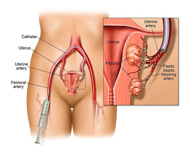
The Process of Prostate Artery Embolization

An IR typically cuts a small hole (incision) on your wrist or upper thigh to access your arteries. Perform PAE. Doctors insert a Foley catheter into an artery inside the urethra and push it towards the prostate to provide a reference point. The radiologist then directs the catheter blood supplying vessels in your prostate.

Once the catheter is in position, the IR injects tiny round particles into the arterial branches through the catheter to reduce or block blood flow. The blockage of these branches is embolization. The radiologist moves the catheter to the other prostate and repeats the procedure.

Embolization deprives the prostate of oxygenated blood, making it shrink and relieve. Hence, improving urinary symptoms after a few days. Once the doctor completes the procedure, they eject the catheter gently from the prostatic arteries.

Embolization lasts about one to four hours, depending on the size and location of arteries in the prostate. Most males feel no pain during the procedure, only a pinch on the wrist or groin at the puncture point.

To reduce anxiety during the process, a doctor may prescribe mild sedatives like Fentanyl. Otherwise, patients usually stay awake during the entire procedure.

Benign Prostatic Hyperplasia (BPH)

As mentioned earlier, Benign Prostatic Hyperplasia is a large prostate gland in older men. BPH can cause urinary problems, but it is not cancer and is not a risk for prostate cancer. If you have the condition, worry less as you have options like PAE to explore.

What are the Advantages of PAE?

PAE is a safe medical procedure for men with BPH. Like many other minimally invasive, PAE has many benefits over traditional surgery.

A year after the procedure, the prostate shrunk by about 30%. As a result, the patient experiences minor urinary symptoms, sparing erectile function, and improves the general quality of life.

Extra benefits of PAE include;

  1. Quick recovery time; one week
  2. Minimally invasive procedure
  3. The procedure is simple and can happen as an outpatient procedure
  4. Low to nil risk of getting erectile dysfunction or retrograde ejaculation
  5. For most patients, there is less need to use a catheter
  6. Less swelling and pain
  7. There is no risk of unrestraint leakage surrounding healthy tissue
  8. The complication rate is less than 1%
  9. A doctor can perform the procedure on a moderate or large prostate gland with no upper size limit
  10. Prostate-specific Antigen levels drop after the procedure
  11. PAE reduces lower urinary tract symptoms (LUTS), e.g., the urgency and frequency of urinating, especially at night. Others are weak urine, straining to urinate, incomplete bladder emptying, and intermittency.

What Are the Risks Of PAE?

Only skilled, qualified and well-trained inter-ventional radiologists should perform PAE. Patients may experience “post-PAE syndrome” for a few days after the procedure. Patients may feel nauseous, vomit, have a fever, abdominal pain, or frequent urination.

Other risks include hematoma at the incision are; blood in urine, semen, or stool. You may also have bladder spasms or an infection of the incision area or prostate.

Please Schedule an Appointment with Us Today

Are you tired of dealing with the side effects of an enlarging prostate? PAE might be the best remedy for your condition. Contact MG Health today and let us answer all questions on prostate health. We have the best medics to diagnose and treat your disorder and walk you through the healing journey.

Reversing PAD

Peripheral artery disease occurs when your arteries have hardened and become narrow because of plaque buildup. The plaque consists of fats and other substances found in your bloodstream, and PAD mainly affects the highways that transport blood to the arms and legs.

When someone has peripheral artery disease, they go through many challenges since the condition makes it harder for their head, legs, arm, and other organs to get enough blood flow.

The fact that all these body parts cannot access enough blood means a likelihood of eventualities like tissue damage and, finally, tissue death. The worst-case scenario is that if this disease does not get the proper treatment, a patient may get a stroke, amputation, heart attack, and kidney disease. Eventually, the patient might die. Again, there is no cure for PAD.

That said, you don’t need to worry. Being diagnosed with PAD is not the end of the road. You don’t have to die, and you also don’t have to suffer from stroke and all the above conditions.

The good thing is that peripheral artery disease can is manageable, and the symptoms can be reversible. It is best to manage the symptoms with modifications in your lifestyle. Alternatively, you can opt for minimally invasive endovascular procedures if the lifestyle changes are not working.

What Lifestyle Changes Can You Make to Treat PAD?

Changing your lifestyle is the easiest way to manage your PAD symptoms; this is a great way to stop the disease from progressing. Here is what you can do:

·      Quit Smoking

Tobacco use is dangerous and a significant risk factor for PAD. Smocking tobacco compresses and damages your arteries. If you have been smoking, it’s high time you considered quitting. When you stop, you will reduce the probability of your symptoms developing further and, simultaneously, lower your odds of cardiovascular disease. 

·      Healthy Diet and Weight

Many people suffering from PAD already have underlying conditions such as high blood pressure, diabetes, and high cholesterol. Embracing a heart-healthy diet is very important in this case because it reduces your risk, and you will have taken care of the other conditions.

Eat a diet low in sugar, avoid processed food, and reduce salt, red meat, and added sugars. Focus on meals with vegetables, fruits, and whole grains. Your diet should have low-fat products, poultry, nuts, seeds, and non-tropical vegetables.

Avoid Certain Over-the-Counter Medicine

Always consult a doctor when you are sick. That is because some medicine could worsen your symptoms instead of curing your diseases. Some contain pseudoephedrine, for example, Advil for cold & sinus and Aleve D Sinus & Cold; those drugs shrink your blood vessels. 

Stop or Reduce Alcohol Intake

Have you ever heard of alcoholic cardiomyopathy3? You might want to stop consuming much alcohol. Cardiomyopathyis a condition caused by too much alcohol consumption, especially when it is long-term. A lot of alcohol will enlarge your heart, and that is irreversible. It will make your heart muscles weak, making the heart stop pumping as it should. Limit your alcohol intake, and your symptoms will not get worse.

Exercise

Exercise is a critical strategy in PAD reversal. Regular physical exercise results in fewer and less severe symptoms. It is possible to assess the success of the PAD treatment by watching the distance in which a patient walks without feeling pain.

Engage in routine walks also. That will help to improve your self-esteem, mood, energy, and sleep. Also, exercising reduces your risk of cardiovascular diseases such as heart attack, stroke, and heart disease.

Take Care of Your Feet

People with PAD may also have diabetes and wounds on the lower part of their legs. That is why foot care is essential for them. Most of these wounds and injuries do not heal. The cause of them not healing is poor blood flow. Such wounds could give you infections.

That is how you take care of your feet:

  • Wash your feet daily and ensure they are dry
  • Moisturize your feet
  • Put on thick dry socks
  • Wear a shoe that fits properly
  • Treat all fungal infections properly
  • Exercise caution when cutting nails
  • Inspect your feet daily to see if there are injuries
  • Have a foot doctor that can treat bunions or calluses

Final Thoughts

When you get diagnosed with PAD, the first thing your doctor will want to do is to control the symptoms you have. They will also want to stop the plaque from building up in your arteries. Lifestyle changes are good enough to meet those goals at times. When that is not enough, the doctor will advise you accordingly.

Radiologic Imaging: Interventional Radiology Techniques

Radiologic imaging is a technic used by interventional radiologists to treat conditions in the vascular system. This technique uses images of your internal body structure to determine the type of treatment that should occur when you have a vascular disorder. Also taken into consideration are pre-procedural planning and imaging.

According to Health Images, diagnostic imaging, or radiology, is a group of procedures involving photos of various body parts. Radiation is used to both make an accurate diagnosis in the fields of diagnostic radiology and interventional radiology.

  • Diagnostic radiology: this technology uses imaging to obtain images inside the body. Diagnostic radiologists then use these images to diagnose injuries and diseases.
  • Interventional radiology: is a medical procedure used by specialists for imaging procedures that obtain pictures from inside your body. The technique is then used to interpret these images to diagnose injuries and diseases and perform life-saving medical functions.

There are different imaging exams. The most common of these include x-rays, ultrasounds, MRI, CT, and PET scans. A radiologist interprets the outcome of imaging tests to evaluate and support a diagnosis.

Types of Radiologic Imaging

There are a number of radiologic imaging types:

  • CT scan (computed axial tomography or CAT scan): This is a non-intrusive medical examination that creates multiple images or photographs of the inside of the body using specialized X-ray equipment and cutting-edge computers.
  • CT lung cancer screening: This screening is a low-dose scan of your lungs used to detect lung cancer.
  • Ultrasound: This medical procedure, also known as sonography, uses high-frequency sound waves and a pc to produce images of the organs, tissues, and circulatory system.
  • Virtual colonoscopy: This medical imaging technique creates 2D and three-dimensional images of the digestive tract that you can view on a video feed using CT scanning and sophisticated computer software.
  • Fluoroscopy: This is a medical procedure wherein the part of the body under analysis undergoes exposure to sustained X-ray beams, which then transfer to a screen that resembles a television to see, in detail, the body part and its movements.
  • Magnetic resonance imaging (MRI) scan: A non-invasive medical examination that takes images from inside your body using a powerful magnet and a computer.
  • X Rays (radiographs): A medical procedure to create images of your internal organs, vertebrae, and tissues on film using invisible electromagnetic energy waves.

Uses of Radiologic Images

Different radiologic imaging technologies are necessary because other conditions affect different body parts.

The most helpful imaging techniques and a few standard procedures are as follows.

Embolization

According to the Center for Vascular Medicine, embolization is a minimally invasive procedure that reduces blood flow to target areas. It protects your tissues and controls problematic blood loss while maintaining the normal flow of blood in the surrounding areas.

Embolism is the definition of a blocked blood vessel. On the other hand, obstructing a blood vessel on purpose for medical reasons is an embolization procedure.

Embolization procedure for:

  • Abnormal bleeding restriction or mitigation
  • Cutting off blood supply to a tumor
  • Repairing a vein’s an artery’s abnormal connections
  • Managing aneurysms
  • Shrinking the size of your veins that are not straight or tangled to lessen pain and swelling

Arterial Revascularization

Non-invasive vascular exams enable doctors to choose the best course of treatment before procedures take place on the legs. Ultrasound is the most frequently used method. Since the leg blood vessels don’t have any overlying organs that could limit accessibility, ultrasound radiologic imaging easily sees the blood vessels in the legs. The process facilitates quick and simple qualification and evaluation.

PVR, or pulse volume recording, also assesses the blood vessels’ efficiency. PVR detects artery disease and assesses treatment response after a procedure. This uses a series of blood pressure cuff metrics at various sites on the legs. Following treatments, PVR tests check to see if any blockages have recurred.

Vein Evaluation

This is a technique that detects the functionality of your veins. It is dependent on the veins that need treatment. For legs, ultrasound is sufficient since there are no overlying organs. For pelvic vein problems (varicocele, May-Thurner Syndrome, pelvic congestion syndrome), cross-sectional radiologic imaging is necessary to examine how badly the pelvic disease has progressed.

Importance of Interventional radiology

For patients who might otherwise require open surgery, interventional radiology lowers costs, recovery times, pain, and risks. As a result, IR has replaced other treatment options for many different conditions.

The range of therapies that IR can successfully deliver is constantly evolving and growing.

Some of the procedures that take advantage of interventional radiology include:

  • Angioplasty- repairing or unblocking blood vessels
  • Stenting- treats narrow and weak arteries
  • Thrombolysis- dissolves blood clots
  • Embolization- blocks the flow of blood to cancer cells
  • Radiofrequency ablation- reduces pain in the nerves
  • Biopsies- used in studying tissues

What Is Interventional Radiology

Interventional Radiology, IR is a generic field within radiology. It is a way to diagnose and treat medical conditions such as cancer without a patient undergoing major surgery.

In IR, a physician does non-surgical procedures (minimally invasive) with the help of imaging tests such as MRI, CT scan, X-Ray, or ultrasound.

In such tests, the radiologist gives the patient a local anesthetic followed by a needle stick. The tests help your doctor to get a picture of how your body looks and may later use needles or tubes to perform a procedure if necessary.

Who Performs Interventional Radiology on A Patient?

An interventional radiologist is a certified medical doctor who performs IR. The interventional radiologist sits for a board exam under the American Board of Radiology, completing the accredited residency program. Later, they complete a fellowship-training program enabling them to work closely with other doctors in patient treatment.

The Working of Interventional Radiology

An interventional radiologist uses ultrasound, MRI, or CT scans to see the inner body parts that need treatment.

Next, a needle goes into your vein with contrast dyes to help get clear images. Some procedures involve using a catheter or wire through a small incision on your body.

Dye follows the blood flow direction to give many details. Then your physician advances a wire through the needle into your blood vessels as they watch the screen.

Alternatively, a physician may perform some procedures through the skin, e.g., liver tumor ablation (“tissue destruction”). Here, the physician inserts a needle through the skin directly into the liver tumor with the help of a CT scan or ultrasound.

The needle connects to an energy source that burns the growth. During the treatment process, you may need sedation to ease pain and help you relax. Often, you won’t need general anesthesia that makes you sleep during the entire procedure.

Interventional Radiology for Cancer

Physicians may use interventional radiology in cancer treatment in many ways. IR can help prevent excessive bleeding during surgery and minimize pain and the side effects of cancer treatment.

A doctor may use it to put chemotherapy and radioactive medicines (cancer-killing therapies) into the cancer cells. Again, a doctor may heat radio waves or electric currents or use ice crystals to kill cancer cells.

Cancer treatment side effects such as the build-up fluids or blood clots can also go through IR.

Examples of Procedures that take place in Interventional Radiology

  • Angioplasty – unblocks or repairs damaged blood vessels to control bleeding
  • Stenting – a physician inserts a tiny mesh coil to unblock blocked arteries
  • Embolization – helps to block blood flowing into cancer cells
  • Needle biopsy. Guided by imaging computers, a physician puts a tiny needle into the patient’s body for tissue biopsy
  • Catheters insertions. Your physician may insert a large vein catheter to administer chemo drugs, hemodialysis, or nutrition
  • IVC filters – where a radiologist will put a small filter into the inferior vena cava, a large vein in the abdomen. The filter traps blood clots that may enter your lungs Gastrostomy tubes- a doctor may put a feeding tube into the stomach of a patient who cannot eat by mouth.

What Are the Benefits of Interventional Radiology?

Interventional radiology has two main benefits. First, it grants your doctor access to internal body parts that require treatment. Again, when your patient is receiving treatment, it is doubtful they will get adverse side effects from the treatment.

IR is beneficial in cancer treatment as chemotherapy or radiotherapy may damage healthy cells. IR helps doctors to focus therapy on tumors and not healthy tissues.

The good news is that, interventional therapy isn’t only for cancer patients. People with kidney disease and cardiovascular problems like narrowed arteries may also benefit from it.

Another benefit is that you do not have to report for admission as an in-patient. So you don’t have to stay to stay overnight in a hospital for an interventional radiology procedure.

How to Find the Best Interventional Radiology Service

To get the best IR services, look for the best physicians with excellent reviews, current practice, and enough experience. Typically, professional companies recognize them as experts in specialized treatment therapies.

Ensure that the doctor is board certified in Vascular, Diagnostic Radiology and IR and Board Certified in Diagnostic Radiology. This assures you of getting quality service from the best physician.

The Importance of Early Screening

When you think of November, a lot comes to your mind. You remember how you get to meet up with your family members, the big meals you share, not to forget the cold weather that has everybody enjoying their warm beverages. That is beside the point. There is a big event that takes place this month known as Movember.

Movember is among the most significant fundraisers held to promote men’s health. This is where men get encouraged to own their health by making early screening a priority, changing their lifestyles, and fostering a caring community. Men engage in growing out mustaches, and that is one of the ways through which they raise funds. 

ECCO empowers men by offering them options that will allow them to take control of their health. Much emphasis is on prostate health. We have our endovascular specialists.

During Movember, our endovascular specialists do not grow out mustaches; instead, they come prepared to handle the concerns of men. They are experts in minimally invasive procedures, allowing men to have options in the treatment process.

Benign Prostatic Hyperplasia

The prostate is one of the male organs. This organ surrounds the urethra. When a man becomes older, his prostate can start enlarging. A prostate exerts much pressure on the urethra.

For this reason, the man begins experiencing problems while urinating. The force also comes with other symptoms that include:

  • Sudden urgent needs to urinate
  • Starting or controlling urination becomes difficult
  • Frequent urination at night
  • Weak or interrupted urination

The symptoms above cause much disruption to a man’s life, which is why early screening is essential.

BPH Treatment

You do not need any treatment when the symptoms are mild. In other instances, the issue cannot go untreated; the patient must take daily medication. The medicine may fail to treat the problem completely.

At that point, there could also be signs of damage from BPH. The patient goes through endoscopic surgery recommended by the doctor, and the surgery takes place through the penis. Alternatively, the patient undergoes traditional surgery to correct the issue.

Going through that kind of surgery can be devastating and uncomfortable for most men. In that case, there is no need to worry. At ECCO, we provide Prostate Artery Embolization, allowing our clients to get a solution in a more dignified manner.

Most of the patients are eligible to go through PAE. Prostate Artery Embolization is an outpatient procedure. It is also minimally invasive, and the doctors do it through a small needle puncture in the patient’s wrist or hip area. It involves moderate sedation, and the period you recover is minimal.

Having gone through PAE, patients report that they have gotten relief from the symptoms in two to four weeks. This process gives patients much psychological comfort, given that nothing gets inserted into the penis.

Are You a PAE Candidate?

We want to walk through the journey of healing together with you. Since most patients are eligible, you will only need to come. Let us begin by performing a complete examination that will help us ascertain that PAE is the most appropriate treatment and then advise you accordingly. 

UFE and Its Effectiveness

As a woman, you play an essential role in maintaining your family’s health. Many women work so hard to keep their families together, but when they have issues with their well-being, they are silent and tend to battle health issues in silence. During recovery after delivering babies, some women have to deal with conditions they find difficult to discuss.

Fibroids are one of those conditions. These are smooth muscular tumors. The tumors form on the interior walls of a woman’s uterus in her childbearing years. 70 to 80 percent of women develop fibroids at the age of 50. Many ladies develop fibroids at 35, making up to 30 percent. We treat uterine fibroids through a method known as Uterine Fibroid Embolization (UFE).

When a woman has fibroids, they are likely to have the following symptoms:

  • Heavy menstrual cycles
  • Constipation
  • Infertility
  • Pelvic pressure
  • Dizziness or fatigue
  • Abdominal enlargement
  • Increased urinary frequency
  • Pain during intercourse

Fibroids are non-cancerous, but most women who have had them have opted for their removal. You should see a doctor immediately when you experience:

  • Pelvic pain that does not go away
  • Overly heavy and prolonged periods
  • Difficulty in emptying your bladder
  • Spotting between periods or bleeding
  • Unexpected low red blood cell count

The doctors have not yet established the cause of fibroids, but these factors are common through research and clinical experience: hormones, genetic changes, extracellular matrix, and other growth factors.

What is Uterine Fibroid Embolization?

UFE is a process professionally handled by an interventional radiologist, and a vascular system specialist. It is a secure and effective procedure with over 85% success rate. UFE brings out a substantial reduction in the size of the fibroids. When done successfully, the symptoms usually improve significantly.

For instance, if you are a woman who has uterine fibroids and you struggle with a heavy menstrual flow, UFE will get you to a place where you have a regular flow. Of all the patients who undergo this treatment process, only 5 to 10 percent may need to redo embolization so that their symptoms can disappear completely.

Uterine Fibroid Embolization is minimally invasive, making it a suitable replacement for myomectomy, medication, or hysterectomy. The specialist inserts a catheter into the legs or wrist during this procedure. Then they inject tiny beads into the blood vessels supplying blood to the fibroids to stop the blood from flowing.

After the end of this procedure, the patient goes home, and the fibroids start shrinking gradually, then they die because they lack blood flow.

What are the Benefits and Risks of UFE?

Through UFE, patients have the advantage of both avoiding surgery and staying in the hospital, and that is because the process takes place in the office. Also, UFE does not cause trauma to the uterus, implying that your fertility status will remain intact, unlike in a hysterectomy.

The fact that you will not go through incisions during the process also means that with UFE, you will not experience complications. If there arises any difficulty, it will get treated so quickly. Significant risks in this process only add up to 1 percent, and the physician thoroughly explains that.

What Happens After Uterine Fibroid Embolization?

Because UFE happens to avoid major surgery, you will not stay in the hospital. Also, you will have a shorter recovery period than other procedures. For most patients, the physician does not give post-procedure restrictions, and you will only need a maximum of ten days to recover.

What you should expect to experience:

  • Fever chills
  • Breakthrough bleeding
  • Chills
  • Low energy levels
  • Pelvic pain

You can liken most of these to a cold

Most patients experience a dramatic reduction of fibroids as soon as the next cycle. With UFE, you will need around three months for the benefits to show, and the fibroids often continue shrinking for the next six to nine months.

ECCO Medical has endovascular specialists trained in minimally invasive procedures. They professionally address problems affecting the vascular system, such as uterine fibroids.

Would you like to learn more about uterine fibroid embolization as an alternative to hysterectomy? Reach out now! At ECCO Medical, physicians are ready to answer your questions and advise you on the best way to go as you fight the fibroids. We are ready to walk with you in your treatment journey and give you the best results.

PAD Signs and Symptoms

PAD stands for peripheral artery disease. It is a condition associated with chronic poor circulation of blood. Often, PAD is interchangeably used with peripheral vascular disease (PVD).

PAD occurs due to the arteries narrowing or blocking due to plaque accumulation. Cholesterol and fats on the arterial walls form plaque, which reduces blood flow. Over time, the situation worsens, and some body parts may lack enough blood.

The specific body parts affected are the feet and legs. For example, there is a close link between PVD and varicose veins, deep vein thrombosis, etc.

The signs and symptoms of PAD differ depending on weight, lifestyle, and age. Most patients suffering from PAD are asymptomatic though some have noticeable symptoms. The observable symptoms are due to the reduced flow of blood to the muscles on the leg. Most individuals experience random pain when walking.

What Are the Risk Factors For PAD?

To mention a few, the risk factors are;

  • Age, especially persons above 50 years
  • Patients with pre-existing conditions such as heart disease, hypertension, diabetes
  • Excessive consumption of tobacco products
  • High blood cholesterol
  • Women in menopause
  • Obesity
  • Lack of physical activity

Note;

When you quit smoking, you may reduce your risk of cardiovascular disease, including PAD.

Watch this video to learn more about the risk factors associated with PAD https://www.youtube.com/watch?v=GkHbvR_6GMk.

Signs of Peripheral Artery Disease

More than 50% of patients with PAD are asymptomatic. But, the most common symptom is muscle activity pain in the calves, hips, or thighs. Muscle movements such as climbing the stairs may cause intermittent pain called claudication. The reason is that the demand for oxygen-rich blood increases with muscle activity.

Blocked leg blood vessels cause pain even at night when you are catching a nap. The leg cramp is dull, and your leg muscles feel heavy, tired, and tight.

More symptoms of peripheral artery disease are;

  • One or both feet or legs feel cold and may have skin color changes, i.e., shiny, blue, or red
  • Discolored toenails
  • Persistent pain in the butts
  • Weak pulses on your feet and legs
  • Loss of hair on the legs
  • The legs feel numb, powerless, and tingling
  • The toes or feet aching or having a burning sensation
  • Heel or ankle wounds that never heal
  • Dead tissues due to insufficient blood flow
  • Impotence
  • Nagging sores on the feet or legs

The Diagnosis of Peripheral Artery Disease

To diagnose PAD, your doctor will ask about the history of your symptoms (if any) and perform a series of medical tests.

Additional medical examinations may include;

Angiography

Angiography is a test that checks for blocked or narrowed blood vessels.

Ankle-Brachial Index (ABI)

An ankle-brachial index is a contrast between the ankle and arm blood pressure. ABI helps in examining blood pressure changes in a given time.

Doppler Ultrasound Flow Studies

The Doppler ultrasound utilizes sound waves of high frequency and a computer to show blood vessels and tissue images. This method measures and checks blood flow – faint or no sound may show blood flow obstruction.

Pulse Volume Recording (PVR) Waveform Analysis

PVR test checks for any fluctuations in the amount of blood in your legs.

Exercise Tests

the doctor performs some of the tests named above pre and post-exercise. This helps the doctors examine patients having symptoms when exercising.

The Medical Treatment for PAD

Are you having any symptoms of PAD? Contact our physicians today. We aim to control the symptoms you have and prevent more complications.

The doctor’s choice of treatment depends on your age, overall health, and response to drugs.

Treatment options include;

  1. Prescribed drugs to improve your blood flow, e.g., antiplatelet agents
  2. Controlling pre-existing chronic medical conditions like diabetes and blood pressure may worsen PAD.
  3. Change of lifestyle like exercising, often quitting smoking, and having a healthy diet.
  4. Angioplasty- the doctor uses a long hollow tube to widen the arteries and increase blood flow.
  5. A vascular surgery. The physician creates a bypass graft in the narrowed or blocked artery area to divert blood flow.

A physician does Angiography before angioplasty and vascular surgery procedures.

A major lifestyle recommendation is that ladies stay away from compression stockings. They hold back the flow of blood in people with peripheral artery disease.

What Complications May Result From PAD?

Complications of PAD often occur due to reduced or no blood flow. They may include:

  • Difficulties walking because of pain
  • Heart attack
  • Stroke
  • Wounds not healing, which may lead to amputation

Stay Vigilant About Your Muscle Health

There is no need to live a life of long-lasting wounds, difficulties walking, or even worse complications. Schedule an appointment with us to check your feet and leg muscles. And meanwhile, eat healthily and do exercise. Stay safe!

Diabetes and Amputation

What is diabetes?

Diabetes is a medical condition relating to your body’s usage of glucose and insulin. There are two forms of diabetes.

·      Type 1 Diabetes

It is also known as juvenile diabetes. Some other people refer to it as insulin-dependent diabetes. According to Mayo Clinic, the reason for the condition is little or no insulin production by the pancreas.

Insulin allows glucose to enter your body cells, leading to the production of energy. It is more prevalent in children but can also be evident in adults. Genetics and some virus lead to the development of the condition.

·      Type 2 Diabetes

It impairs your body’s ability to control and utilize glucose. It is a chronic condition. It results in excessive circulation of sugar in your bloodstream. Over time it results in cardiovascular, nervous, and defense mechanisms disorders.

There are essentially two issues at play with type 2 diabetes. Either your pancreas doesn’t produce enough insulin, or your body cells’ reaction to insulin is poor, resulting in low sugar absorption. It is more prevalent in adults than children. Though it has no cure, exercise, weight loss, and healthy eating can help you manage the condition.

According to the Center for Disease Control, people with diabetes have increased from 26 Million in 2010 to around 29 million. Of these, one in four people is unaware they have the condition.

CDC continues to state that upwards of one in three American adults, or an additional 86 million people, have prediabetes, a condition in which blood sugar levels become elevated but not high enough to fall under type 2 diabetes.

Symptoms of diabetes include:

  • Frequent urination
  • Increased thirst
  • Fatigue
  • Presence of ketones in your urine
  • Unexplained weight loss
  • Blurred vision
  • Frequent infections in the gums, skin, or vagina

Diabetes and Amputation

Inadequate insulin results in the accumulation of sugar in the bloodstream. This leads to your blood vessels becoming rough and inflexible, damaging them. As a result, the building up of sticky plaques on the damaged blood vessels.

The condition is called atherosclerosis and it occludes blood flow. Occlusion mainly affects the tiniest blood vessels in the body. These vessels exist in the extremities. Consistent high blood sugar accumulation in the body mainly affects the most peripheral extremities.

Restricted blood flow to your toes, feet, and legs prevents them from getting sufficient oxygen, nutrients, and healing cells. This condition is peripheral artery disease (PAD). They become more vulnerable to nerve damage, infection, and inadequate wound healing as a result.

Since you have no nerve sensation in the injury, you might not notice the wound for a while. This is neuropathy. Chronic wounds may result in tissue damage or infections. When the tissue damage is too severe, amputation is the only solution.

Preventing Peripheral Artery Disease

Early detection of PAD can help avoid amputations. Controlling your blood sugar levels and adhering to a proper diet can help prevent peripheral artery disease.

Additionally, check your A1C blood sugar levels and take app medicine as your doctor directs. Regular physical activity, abstaining from smoking, and avoiding or controlling other health conditions like blood pressure and high cholesterol are additional ways to prevent PAD.

It is vital to watch out for PAD to avoid amputations. Symptoms of the peripheral artery disease include:

  • Leg or pain in the foot which increases during walking or other activities
  • Leg numbness and or weakness – one foot, when compared to the other foot and the rest of the body, may feel colder
  • Slowed nail and hair growth
  • Presence of sores that take long to heal
  • Skin discoloring on your feet or your legs
  • A weak pulse when you visit your doctor

Other Risks of Diabetes

Atherosclerosis is the hardening of arteries. Previously, scientists believed that too much cholesterol buildup in arteries causes atherosclerosis. Science Daily states that cardiovascular diseases happen when blood vessels are totally blocked.

Today, most people concur that a body’s immune environment causes the threat of heart attacks with respect to fatty accumulation than the buildup itself.Diabetes doubles the risk of a heart attack or stroke because it increases the inflammation associated with atherosclerosis.

Other risks associated with diabetes include:

  • Heart diseases
  • Stroke
  • Vision loss
  • Kidney failure
  • Premature death

Type 1 diabetes is unpreventable. However, you can prevent prediabetes and type 2 diabetes by maintaining a healthy lifestyle. Regularly checking your blood sugar levels allows you to catch diabetes early and manage it.

Uterine Fibroid Embolization (UFE)

Uterine Fibroid Embolization (UFE)

Do you have painful fibroids but aren’t ready for a hysterectomy? You’ve got options.

Uterine fibroids are most common in women in their 30s.

What are uterine fibroids?

Uterine fibroids are firm, muscular growths in the uterus that can range from undetectable by the human eye to the size of a melon. They can be single or in clusters. Some never cause symptoms and remain undetected, while others disrupt the woman’s life. You may never know you have a fibroid until you have symptoms, but if you have symptoms, you dread the monthly pain and heavy bleeding.  

As many as 50% of women in their reproductive years have fibroids.

Fibroids are more than a “bad” period

It’s hard to explain to anyone who hasn’t experienced it. Fibroids do more than make your period “worse.” They cause a range of symptoms that can depend on the location, number, and size of the fibroids. For some women, a heavy period is just the beginning. Excessive bleeding, extra-long periods, pelvic pain, frequent urination, constipation, back and leg pain – fibroids can be debilitating.

As many as 80% of women develop fibroids by age 50.

What is Uterine Fibroid Embolization (UFE)?

UFE addresses the fibroid problem without removing the uterus. UFE is a minimally invasive procedure done as an outpatient procedure under mild IV sedation. The physician uses X-ray guidance to thread a catheter through a small incision in the groin and guide it into the uterus. Then, an injection of microspheres cuts off the blood flow to the fibroids, essentially starving them. With their supply cut off, they shrink, relieving your symptoms.

Interventional radiologists have performed UFE to treat uterine bleeding since the 1970s. It’s a safe, effective, proven method of treating symptomatic fibroids.

UFE relieves fibroid symptoms without the complications, side effects, recovery time, and decreased sexual function associated with hysterectomy.

UFE completely or significantly relieves fibroid-related symptoms in almost 90% of women who try it.

How soon will you feel relief?

Symptom relief is noticeable within a few days. The fibroids continue to shrink for several months, and by six months post-UFE, your symptoms will have stabilized.

Women who undergo UFE can typically go home the same day and can expect to resume normal activities in one to two weeks.

Many women who had a hysterectomy for fibroids didn’t know they had another option.

You’ve got options! If you’ve been told you need a hysterectomy for your fibroids, call for a second opinion. Many hysterectomies simply aren’t necessary when UFE is an option.

If you’re living with the debilitating symptoms of fibroids, call us at +1 262-656-2508 for a consultation. We’d be happy to discuss your options and whether UFE is right for you.

Venous Insufficiency

Did you know that a single dysfunction of your veins can lower the quality of your day-to-day life?

The heart relies on veins to keep moving. Consequently, your legs have veins containing valves that let blood out but not backward. Failure of these valves to function efficiently causes blood to pool in your legs, leading to venous insufficiency.

What is Venous Insufficiency?

Venous insufficiency is a condition that occurs when veins in the lower legs are unable to pump blood back to your heart effectively. This can cause blood to pool in your legs, leading to several symptoms.

For example, venous insufficiency can cause the legs to feel tired, achy, and heavy. It could also contribute to leg swelling, varicose veins, and other problems.

While venous insufficiency is not usually life-threatening, it can be uncomfortable and debilitating.

Is there a Cure for Venous Insufficiency?

There is no promising cure for venous insufficiency. But there are treatments that can help manage the symptoms and improve your quality of life.

What are the Signs and Symptoms of Venous Insufficiency?

There are several signs and symptoms associated with venous insufficiency. The most common symptoms include:

  • Swelling in the legs

Leg swelling is a leading consequence of blood pooling in the legs. This causes the legs to become tender. Your legs may swell after standing or sitting for a long time.

  • Cramping in the legs

Pooled blood in your legs may cause leg cramps that are extremely painful. The cramps may also manifest in violently contracting muscles. Venous cramps occur in the calf.

  • Varicose veins

Increasing blood pressure in your veins eventually leads to varicose veins. Varicose veins are a condition in which the veins become enlarged, lumpy, and twisted. They can occur anywhere in the body but are most common in the legs and feet. Varicose veins can be painful and can cause ulcers.

  • Skin ulcers

Venous ulcers occur due to the high pressure in veins in your lower leg. This pressure usually is due to blood in these veins flowing toward your heart. At this point, you may have ulcers or sores on your legs that are slow to heal.

  • Blood clots

The skin around your legs may become darker than usual. This implies that the forward blood flow through the veins is obstructed.

  • A feeling of heaviness in the legs

You may feelheaviness or pain in your legs, especially after standing or sitting for long periods. You may also notice that your legs feel exhausted more often.

  • Dry and itchy skin

Blood builds up in damaged vessels, eventually leaking into the skin. The leaking blood vessels may prevent enough oxygen from getting to your skin. With time, the skin over these veins becomes red and itchy. 

  • Dark purple or blue color on the skin surface

Dark purple or blue color on your skin surface occurs when blood pools in the veins.

If you have any of these symptoms, it’s vital to see a doctor so you can get a proper diagnosis and treatment.

What are the Risk Factors for Venous Insufficiency?

There are several risk factors for developing venous insufficiency. These include:

Age: Venous insufficiency is more common in older adults. As you become older, your blood vessels may lose elasticity or weaken, which contributes to venous insufficiency.

Gender: Unlike men, women are at a higher risk for venous insufficiency. This is due to the effects of pregnancy and menopause on the vascular system. Pregnancy, for instance, may put pressure on the legs contributing to venous insufficiency.

Family background: Family history is another important risk factor. The condition can be hereditary.

Lifestyle choices: Poor lifestyle choices may manifest in obesity and a sedentary lifestyle. Being overweight and obese or even sitting or standing for long may increase pressure on the legs, contributing to venous insufficiency.

Phlebitis: Phlebitis occurs when a blood clot forms in a vein. This condition can cause the vein to become inflamed, leading to venous insufficiency.

Smoking history: Smoking damages the veins and makes it more difficult for them to function correctly.

History of blood clots: When a clot forms, it can block blood flow through the veins. This can cause the veins to distend and weaken, leading to venous insufficiency.

History of deep vein thrombosis (DVT): DVT is a condition that occurs when a blood clot forms in a deep vein, usually in the leg.

Week blood vessels: Weak or damaged blood vessels cannot pump blood as effectively as healthy veins. Weak blood vessels can result from certain medical conditions or even pregnancy.

Veins in Our Body Have an Important Role

When the veins in our body cannot pump blood effectively, blood begins to pool in the vessels. This can cause several problems. It may cause pain, swelling, blood clots, ulcers, and even varicose veins, among other circulation problems.

Thankfully, there are treatments available that can help to improve veins’ function and prevent blood pooling.

What are the Treatment Options for Venous Insufficiency?

The treatment options for venous insufficiency include:

  1. Varicose Vein Procedures

Treatment for venous disease may vary by diagnosis. But varicose veins procedures can help with the situation.

Invasive procedures can improve blood flow to the heart. The procedure closes the damaged vein and reroutes blood to the healthier veins.

Another procedure involves injecting non-formulated medical adhesives to close the damaged vein.

Additional treatment options involve using heat to close off the sick veins. These include:

  • Ablation (RF ablation)
  • Endovenous Laser Ablation (EVLT)
  • Regular Vein Treatments

Regular vein treatments such as sclerotherapy can help. So, what is sclerotherapy?

Sclerotherapy is a medical treatment for varicose and spider veins. It involves injecting a chemical solution into the veins. This injection causes the vein to shrink and disappear. Sclerotherapy is safe and effective for reducing the appearance of varicose veins.

  • Surgery

In more severe cases, surgery may be necessary to remove the damaged veins.

  • Non-surgical Treatment Options

Other than medical treatment, there are several measures you can take at home to improve venous insufficiency. These may include lifestyle changes such as:

  • Wearing compression stockings to compress the damaged veins by applying pressure to the legs. This improves blood flow and reduces swelling in the legs.
  • Wearing loose-fitting clothes
  • Elevating your legs when sleeping or sitting
  • Avoid sitting or standing for long periods if you have varicose veins
  • Practicing healthy eating habits and exercising regularly
  • Avoiding smoking

Working with a qualified healthcare professional is essential for whichever treatment option you choose. This will ensure you get the best possible results in managing venous insufficiency.